インスリン受容体 IR: 構造、機能など
UBC/protein_gene/i/ir
このページの最終更新日: 2026/04/09- 概要: インスリン受容体とは
- インスリン受容体の構造
広告
概要: IRS とは
インスリン受容体 (IR; insulin receptor) は、インスリン のシグナルを伝える分子の一つで、細胞膜に存在しインスリンと結合する。
インスリンが IR に結合すると、IR の細胞内ドメインのチロシンキナーゼが活性化し、IR を自己リン酸化する。IRS はこのリン酸化チロシンキナーゼを認識し、IR に結合する。結合後は、IR のチロシンキナーゼによって IRS のチロシンもリン酸化され、ここに複数のタンパク質が結合し、シグナルをさらに下流に伝える。
IRS の下流には、次のような分子があることが知られている。
広告
「あとがき」で当サイトを参考にしたと書いてくれているラノベです。Kindle Unlimited で読めました。ストーリーと文章が良く、面白かったです。
インスリン受容体の構造
ヒトのインスリン受容体は、22 のエクソンから成る 1 回膜貫通型受容体である。エクソンと各ドメインを示した良い図があったので載せておく (1)。
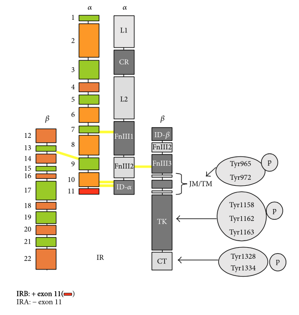
Structure and phosphorylation residues of IR, IGF-IR, and hybrid receptors. (a) Schematic representation of IR. (Left) α chain: exons 1–10 (IRA) or exons: 1–11 (IRB) and β chain: exons 12–22. (Right) Domains of IR: L1, large domain rich in Leu; CR, domain rich in Cys; L2, large domain 2; Fn, fibronectin III type domain; TM, transmembrane domain; JM, juxtamembrane domain; TK, Tyr kinase domain; CT, C-terminal domain. In the JM, TM, TK, and CT domains, Tyr phosphorylation residues are indicated.
広告
References
Escribano et al. 2017a. The role of insulin receptor isoforms in diabetes and its metabolic and vascular complications. J Diabetes Res 2017, 1403206, 2017.
コメント欄
サーバー移転のため、コメント欄は一時閉鎖中です。サイドバーから「管理人への質問」へどうぞ。