結合組織と細胞を区別する MT 染色: 原理、方法など
UBC/experiments/tissue_staining/mt_staining
このページの最終更新日: 2025/11/23広告
概要: MT 染色とは
日本語では「マッソントリクローム染色」と表記する。以下の 3 つの染色の組み合わせである (1)。
- ヘマトキシリンで 核 を染める。
- 拡散速度の大きい分子 (酸性フクシン、ポンソーキシリジン) で細胞を染める。
- 拡散速度の小さい分子 (アニリン青) で組織構造が荒い結合組織を染める。
これによって細胞と結合組織の染め分けが可能になる。組織に結合組織が入り込んでくる筋ジストロフィー (参考: ジストロフィン) 研究などの分野でよく用いられる。
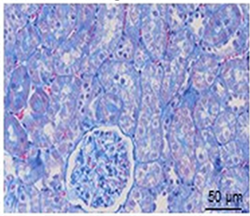
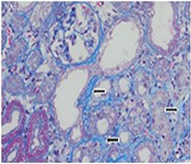
腎臓の MT 染色像
図 (Ref. 2) は 腎臓 の MT 染色例である。論文では H&E 染色 および PAS 染色結果とともに示されているが、ここでは MT 染色のみを載せておく。
これは adenine および chrysin が腎臓の繊維化を引き起こすという論文で、Control に比べて処理群では青色の結合組織がはっきりと認められる。
|
Control 
|
Fibrotic (adenine + chrysin 処理) 
|
広告
References
- マッソントリクローム染色. Ppt file: Last access 2018/03/15.
Ali et al. 2015a. Ameliorative effect of chrysin on adenine-induced chronic kidney disease in rats. PLoS ONE 10, e0125285.
Ali et al. (2015a) is an open-access article distributed under the terms of the Creative Commons Attribution License, which permits unrestricted use, distribution, and reproduction in any medium, provided the original author and source are credited. Also see 学術雑誌の著作権に対する姿勢.
コメント欄
サーバー移転のため、コメント欄は一時閉鎖中です。サイドバーから「管理人への質問」へどうぞ。
