高分子の炭水化物を染める PAS 染色:
原理、プロトコール、写真など
UBC/experiments/tissue_staining/pas_staining
このページの最終更新日: 2026/04/09- 概要: PAS 染色とは
- 実際の染色例
- 粘液細胞は強く染まる
広告
概要: PAS 染色とは
PAS 染色 (Periodic acid-Schiff staining) は、グリコーゲン、糖タンパク質、プロテオグリカン、セルロースなどの
まず、過ヨウ素酸 periodic acid (図, public domain) が糖を酸化してアルデヒドを生成する。Wikipedia には「グルコース を選択的に酸化」と書かれているが、これは間違いである。
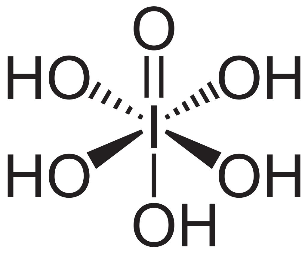
次に、シッフ試薬 Schiff reagent によってアルデヒドを赤紫色に染色する。その結果、グリコーゲン、糖タンパク質などの
実際の PAS 染色例
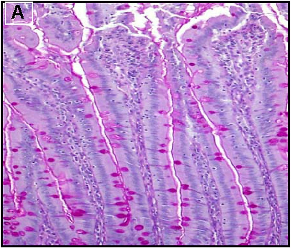
粘液細胞は強く染まる
下の図 (1) は、小腸絨毛の PAS 染色結果を示している。粘液を分泌する杯細胞 goblet cells が染色されている。これは、多糖が粘液の主要成分であるためである。
 広告
広告References
Wang et al., 2015a. Human ghrelin mitigates intestinal injury and mortality after whole body irradiation in rats. PLoS ONE 10, e0118213.
Wang et al. (2015a) is an open-access article distributed under the terms of the Creative Commons Attribution License, which permits unrestricted use, distribution, and reproduction in any medium, provided the original author and source are credited. Also see 学術雑誌の著作権に対する姿勢.
コメント欄
サーバー移転のため、コメント欄は一時閉鎖中です。サイドバーから「管理人への質問」へどうぞ。