バルビアニ小体とは: 定義、特徴など
- 概要: バルビアニ小体とは
広告
概要: バルビアニ小体とは
バルビアニ小体 Balbiani body とは、フランスの科学者 Balbiani が 1800 年代にクモで報告した構造である (1)。卵細胞 oocytes にみられる構造で、ミトコンドリア、小胞体、ゴルジ体、RNA、germinal granules などに富む。
このページ では a membraneless ball of mitochondria, other organelles and proteins that breaks apart when the oocyte begins maturing into an egg と書かれている。
ショウジョウバエ、カエル、魚、マウス、ヒトの卵細胞などで報告がある (1)。Mitochondria cloud と呼ばれることもあるようだ。
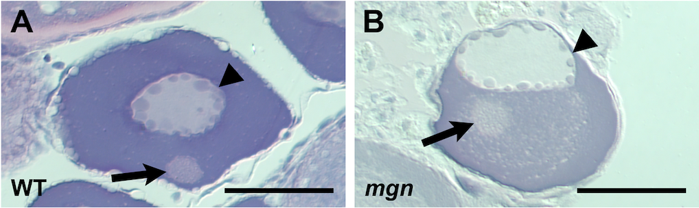
下の図 A (文献 2 より) はゼブラフィッシュの Stage I oocyte の H & E 染色 像である。中央の矢尻で示された部分が核 nucleus、パネル下の矢印がバルビアニ小体。これぐらいのサイズで見える。
B は mgn という遺伝子の変異体で、核とバルビアニ小体の位置が変わってしまう。
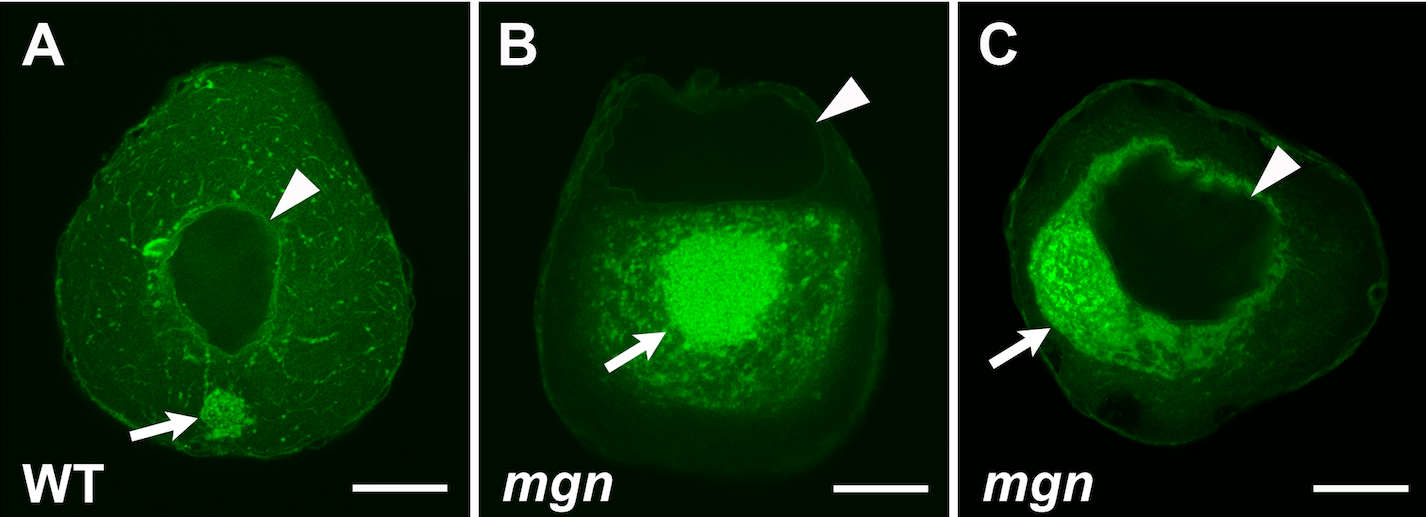
下の図も同じ論文 (文献 2) から。蛍光染色像で、同じく矢尻が核、矢印がミトコンドリア。mgn 変異体ではバルビアニ小体の肥大が認められる。
広告
References
Schisa 2012a. Chapter seven - New insights into the regulation of RNP granule assembly in oocytes. Int Rev Cell Mol Biol, 295, 233-289.Gupta et al. 2010a. Microtubule actin crosslinking factor 1 regulates the Balbiani body and animal-vegetal polarity of the zebrafish oocyte. PLoS Genet, 6, e1001073.
Gupta et al. (2010a) is an open-access article distributed under the terms of the Creative Commons Attribution License, which permits unrestricted use, distribution, and reproduction in any medium, provided the original author and source are credited. Also see 学術雑誌の著作権に対する姿勢.
コメント欄
サーバー移転のため、コメント欄は一時閉鎖中です。サイドバーから「管理人への質問」へどうぞ。