光受容細胞: Photoreceptor cell
2021 年 11 月に URL 変更、リダイレクトしました。
広告
概要: 光受容細胞とは
図は Reference 3 より転載したもので、C に
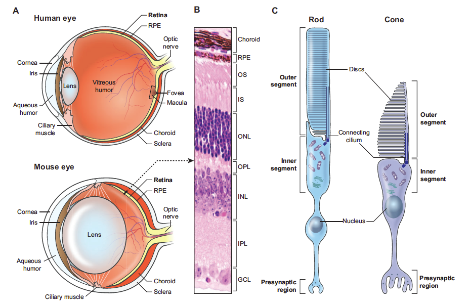
桿体細胞および錐体細胞には、次のような特徴がある。
桿体細胞 |
ロドプシンを含み、感度が高いが色を認識できない。 |
錐体細胞 |
色を認識できるが、多くの光量が必要である。 |
disc 部分は常に RPE に消化されており、毎日新しい disc が供給されている。10 - 15 日ほどで disc 全体が入れ替わる。
なお、網膜で受け取った信号は、まず視床の神経核 lateral geniculate nucleus (LGN) へ運ばれ、さらに primary visual cortex へ運ばれる (4I)。
Primary visual cortex (V1) からは inferiol parietal lobule へ投射があり、これは dorsal stream (where system,

広告
References
ベアーほか 2007a. 神経科学 脳の探求. 西村書店. Amazon link.Meier 2014a. Collinear features impair visual detection by rats. J Vis 11, 1-16.Veleri et al. 2015a (Review). Biology and therapy of inherited retinal degenerative disease: insights from mouse models. Dis Model Mech 8, 109-129.Butler et al. 2006a. Visual white matter integrity in schizophrenia. Am J Psychiatry, 163, 2011-2013.
Veleri et al. 2015a was published as an Open Access article distributed under the terms of the Creative Commons Attribution License, which permits unrestricted use, distribution and reproduction in any medium provided that the original work is properly attributed. Also see 学術雑誌の著作権に対する姿勢.
コメント欄
サーバー移転のため、コメント欄は一時閉鎖中です。サイドバーから「管理人への質問」へどうぞ。